What is Lymphedema?
Lymphedema is a disorder where the protein rich fluid of the interstitial space cannot be cleared by lymphatic system. The reasons may be absent or fewer lymphatics by birth, blockage of the lymphatic channels secondary to trauma, filariasis, cancer, radiotherapy.
What is lymphatic system?
Just like in cardiovascular system where the blood is pumped from the heart and is circulated over the body in arteries and vein, lymphatic system is also a network of fine delecate channels spread throughout the body. The basic role of lymphatic system is to collect fluid laden with proteins, fat and waste products (lymph) which is constantly leaking out of the porus vascular system. This excess fluid is filtered and the bacteria and foreign bodies are removed at the lymph nodes. The fluid is returned back in to the venous system and gets back into the circulation.
How to identify it?
In the beginning patients will only have swelling of their feet or hands which resolves with elevation. With progression of the disease, the swelling does not reduce even with elevation of the limb. In later stages the skin becomes thick and hard. There can be lymphatic fluid leakage from wounds or cracks in the limb. Recurrent infections and skin breakdown can cause significant debilitation.
What are the symptoms that should draw your attention?
Even in the first stage, swelling of feet or hands that reduce on elevation needs attention. The reason for this swelling needs to be investigated as it could be from various medical causes like malnutrition, reduced proteins, liver, kidney or cardiac diseases. Classic lymphedema due to lymphatic obstruction is seen in filariasis or sequelae of cancer and radiotherapy.
In the second stage swelling increases and limb elevation will not reduce it. Further it can progress to the third stage where the skin is hard, pigmented and fissures develop. Infections are more likely at this stage.
At any stage, it is advisable to consult a doctor specialising in lymphatic surgery. With advancements in understanding of the lymphatic physiology and the pathological process, today there are various treatment modalities available and the results have been encouraging.
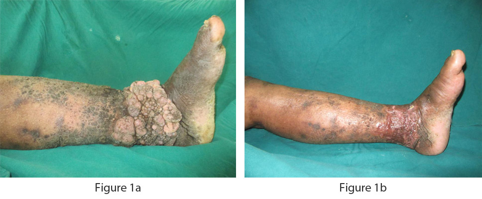
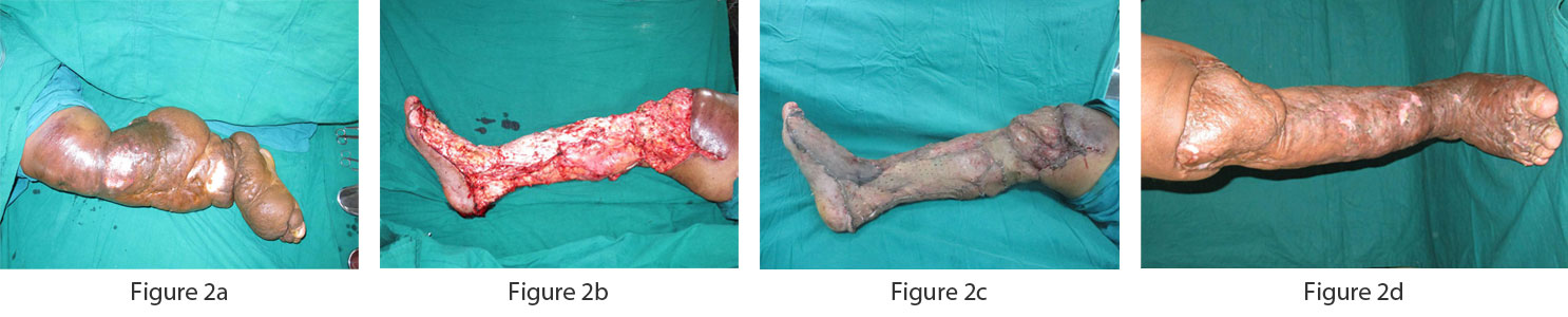
In the late stage called elephantiasis, the limb is very large, heavy, and difficult to lift or mobilize. It will be laden with nodules, papillomas, fissures and may be discharging a clear fluid called lymph. Such patients will need multimodal treatments and staged debulking surgeries.
Is it too late to get treatment?
Now is the best time. At whatever stage you are, the treatment can be started. For a motivated patient, there is good chance of improvement.
What tests are required to diagnose lymphedema?
A clinical history and examination are usually enough. A lymphoscintigraphy or MR Lymphangiography can be done to map the lymphatic channels in advanced cases and also for planning lymphatic surgeries.
How is lymphedema measured?
LIMB MEASUREMENTS
Volumetric displacement of water
Although the method appears clumsy, it is the cheapest of the methods which can be used to measure the limb volume. It works on the Archimedes principle, and the volume of displaced water is the limb volume.
The problems faced are need for a large container, it may not always be able to immerse the whole limb. Also with patients coming with skin complications and raw areas, it appears impractical to measure volume by this method.
Perometry
It uses infrared technology and computer algorithms to measure the limb circumference. When compared to bio-impedence spectroscopy, perometry cannot give the volume of each compartment in the limb like muscle, interstitial compartment etc. However it is a helpful tool to detect lymphedema before it becomes clinically evident and made out by tape measurements.
The perometer works with two LEDs illuminating the limb from a frame. The lights are perpendicular to each other. The frame needs to be moved over the whole extent of the limb. The data is used by a software which gives the limb measurement.
Bio-impedence spectroscopy
It has the advantage over peromety in its ability to map the fluid compartments. Total body water, ECF and interstitial fluid can be calculated. Various frequencies of electrical current passed through the body will be assessed by computer algorithms. Lower frequencies do not penetrate the cells while higher frequencies do. The resistance through various tissues is the basis for the readings obtained.
Mapping Of The Lymphatics
Lymphoscintigraphy
Techicium 99 sulphur colloid or micro albumin are used for lymphoscintigraphy. Tracer is injected in the 1stwebspace of the foot or second webspace in the hand. Clearance of the tracer is then mapped by a Gamma camera – immediately after tracer injection and at intervals of half hour, one hour and delayed image at 4 hours. If lymphatics are absent, there is no movement of the tracer. In case of paucity of lymphatics, the tracer is seen along the path of these lymphatics. The image will show dermal backflow in case of obstruction.
Mr Lymphangiogram
High resolution images of the lymphatics can be obtained and three dimensional reconstructions done. Heavy T2 weighted sequence is used for knowing the extent of lymphedema, while T1 weighted sequence with fat saturation is used for lymphatic visualization
Pathological lymphatics are visualised as tortuous and beaded, there could be dermal backflow in proximal obstruction and honeycombing in case of collateral transport. The advantage with MRL is the visualisation of nearby veins for planning LVA, mapping of the lymph nodes. MRL can also be used postoperatively for mapping the patency of the lymphatic/lympho-venous anastomoses.
Fluorescent Lymphangiography
Indocyanine green injected in the web space is mapped using a SPY camera. It is a real time imaging method which can be used just before surgery to map the lymphatics. Post LVA or lymphatic reconstruction the patency in the anastomoses can also be checked. Although there are so many pros, the cons being required to buy expensive SPY camera; difficulty to image the lymphatics following diffusion, which usually occurs after an hour of the contrast injection are notable problems. The ease of performing the test real time inside the operation theatre safely and accurately scores over the other methods.
Treatment Strategies
Are there non-surgical options?
Yes. Comprehensive decongestive therapy (CDT), 4 layer bandaging, graded compression stockings, pneumatic pumps help reduce the accumulated fluid and aid control of lymphedema to a great extent in early stages. They may be continued post-surgery to sustain the improvement.
Comprehensive Decongestive Physical Therapy
It is not to be confused with massage techniques and manual lymphatic drainage. The principle behind CDPT is not just to move the fluid proximally, but to stimulate the intrinsic mechanisms to start working. Exercises are done focussing on pumping the lymph. Another important aspects is skin care, and prevention of infection which complicate the problem.
Multilayer compression garment is applied. For the patient to be able to meet the therapist regularly, a pneumatic compression device can be used. Patients can do it in the comfort of their homes.
What are the common surgeries?
Surgery is divided into two types.
Type I - Reduction surgery – Fig. 1
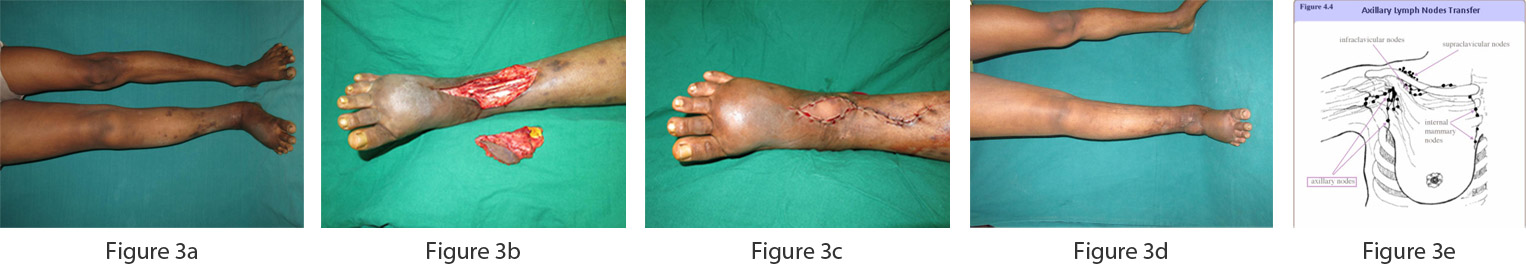
Type II – Lympho Modulating Surgery
DOs and DONTS FOR LYMPHEDEMA PATIENTS
DOs
DON’TS
Patients must understand that lymphedema is a treatable condition and must forego any stigmata. They should not shy in seeking early treatment.
It has not shown to have better clearance of lymph than a CDT. Hence CDT by a trained physiotherapist is an important.
Lymphatic leakage can be problematic socially with the patient’s leg emanating foul smell, while each infection irreversibly worsens the skin condition.
Immediate admission to a hospital and intravenous antibiotics are a must in case of lymphangitis. Further, a drainage or debridement may be necessary.